
Our periodontal gallery contains images of the types of cases that might necessitate periodontal treatment. The purpose of this gallery is to display the types of results that can be achieved through periodontal surgery.
Periodontal Gallery Case
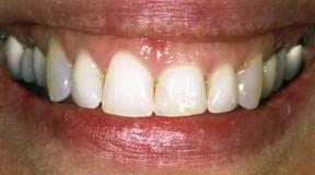
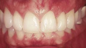
This patient was unhappy with her gummy smile. The gum tissues covered up too much of the teeth, making the teeth look too short.
A crown lengthening procedure was performed, which evened the height of the gum tissue across the front teeth. The result was dramatic and the patient was quite pleased.
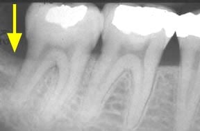
The yellow arrow above is pointing to an area where this patient has suffered some bone loss. The dark area indicates that the bone is not as dense and without treatment the prognosis for this tooth would be poor.
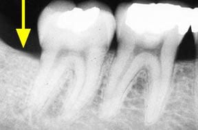
A bone graft procedure was performed and the yellow arrow above is pointing to the regenerated bone. This area is much denser with bone than the same area in the image to the left. The prognosis for this tooth is now excellent.
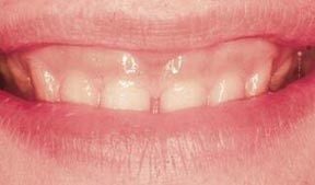
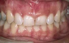
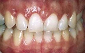
This is another case of a gummy smile. The gum tissue covered up too much of the teeth, which caused them to look short.
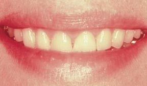
A crown lengthening procedure was performed, which evened the height of the gum tissue across the front teeth. This type of procedure typically leads to patients smiling with more confidence... and much more often!
What is unusual about this case is the unevenness of the tissue height in the two front teeth.
A crown lengthening procedure was performed to make the height of the tissue consistent on both sides of the upper teeth.
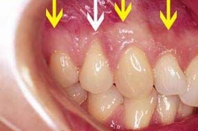
This patient is missing a lot of gum tissue around the neck of the tooth with the white arrow. In this case, it is necessary to do a gingival graft, which requires taking tissue from a site right next to the area missing the tissue or from the roof of the mouth, which contains plenty of healthy tissue. The yellow arrows are pointing to the line of healthy attached tissue, which is needed for the ongoing health of the teeth and underlying bone. You can see how much space there is between the yellow arrows and the neck of the tooth and how little space there is between the white arrow and the neck of the tooth.
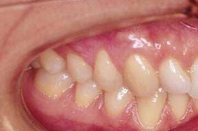
This is the result of a gingival graft, which shows the presence of pink and healthy attached gingival tissue around the neck of the tooth. The prognosis for this tooth is excellent and the procedure took one appointment.
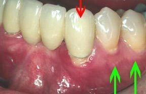
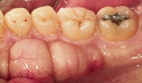
The tissue at the gumline of the tooth with the red arrow is not only very red but it is missing what is called the "attached tissue." Without this tissue, bone loss is likely to occur. This patient does have attached tissue on the other teeth, from the neck of the tooth to the line that the green arrows indicating.
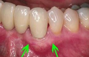
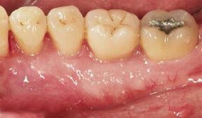
Tissue was taken from the roof of the patient's mouth and grafted to the area shown by the green arrows. Compare the image to the right with the image above. The tissue is much pinker and, therefore, healthier in the above image. The treatment, which took one appointment, should mean that this tooth will be of service to this patient for many more years to come.
The large mass that you see above is simply an overgrowth of bone. It occurs in about 20% of the population. It only needs to be removed for cosmetic reasons, if it interferes with speech, or if a removal appliance such as a denture needs to be made.
This was a one appointment procedure, which was performed because the large overgrowth of bone was affecting speech and eating.