Request an Appointment

Toggle navigation
  • Facebook
  • blog
2607 Davie Boulevard Ft. Lauderdale, FL 33312(954) 587-7111
  • Home
  • About Our Team
    • Meet Our Doctors
    • Meet Our Team
  • Office
  • Dental Services
    • Cleanings and Prevention
      • Bruxism and Mouthguards
      • Fluoride Treatments
      • Sealants
    • Cosmetic Dentistry
      • Bonding
      • Bridges
      • Crowns
      • Dentures
      • Fillings
      • Implants
        • Single Tooth
        • Multiple Teeth
        • All Teeth
      • Inlays and Onlays
      • Lumineers
        • Lumineers FAQ
        • Before and After
        • Perfect Smile
        • Lumismile
      • Teeth Whitening
      • Veneers
    • Gum and Bone
      • Bone Grafting
      • Cleanings and Prevention
      • Crown Lengthening
      • Frenectomy
      • Gum Tissue Graft
      • Perio Plastic Surgery
      • Ridge Augmentation
      • Scaling and Root Planing
      • Sinus Lift
    • Oral Surgery
      • Fractured Teeth
      • MDI Mini Dental Implants
      • Oral Cancer Exams and Biopsies
      • Pericoronitis
      • Wisdom Teeth
    • Invisalign
    • Root Canal
      • Knocked Out Tooth
      • Root Canal FAQ
      • Root Canal Surgery
      • Traumatic Injuries
    • Implants
    • TMJ
  • Smile Gallery
    • Bonding Gallery
    • Crowns Gallery
    • Dentures Gallery
    • Fillings Gallery
    • Implants Gallery
    • Orthodontic Gallery
    • Periodontal Gallery
    • Root Canal Gallery
    • Six Month Smiles Gallery
    • Veneers Gallery
    • Whitening Gallery
  • technology
    • DEXIS
    • Biolase
    • Vatech
    • Straumann
    • Orascoptic
    • Opalescense
    • Treswhite
    • Under Armour Mouthguards
      • Under Armour FAQ
    • Invisalign
      • Invisalign Teen
  • New Patients
  • Contact Us

Root Canal Gallery

Online Dental Education Library

Our team of dental specialists and staff strive to improve the overall health of our patients by focusing on preventing, diagnosing and treating conditions associated with your teeth and gums. Please use our dental library to learn more about dental problems and treatments available. If you have questions or need to schedule an appointment, contact us.

If the nerve of a tooth gets infected, it can be extremely painful. The goal of root canal treatment is to relieve pain, and restore your smile!

Case   of  

Root Canal Gallery Case  

Root Canal Before

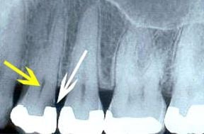
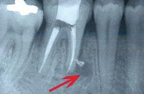
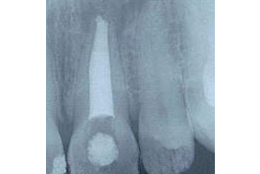
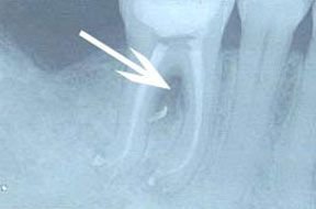
A root canal procedure involves removing diseased tissues (indicated by the yellow arrow), which contain the nerve and blood supply of a tooth, and replacing it with a rubber-type material. The area that is filled is called the "root canal." The root canal of each tooth is very different. The reason this tooth needed a root canal was due to decay (indicated by the white arrow) entering the pulp.

Root Canal After

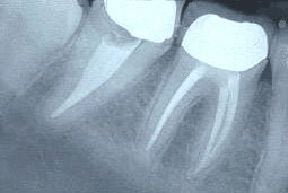
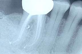
This is what the completed root canal looks like on an x-ray. This tooth had three canals, which you can see by the white filling material in the root portion of the tooth.

Root Canal Before

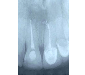
This tooth has what is called an "accessory canal," which is a canal that splinters off in an unexpected direction. This tooth had to be retreated due to the first root canal failing, which is evident by the dark area, which is where the arrow is pointing. Upon retreatment, the accessory canal was found and filled, and the prognosis for this tooth is good.

Root Canal After

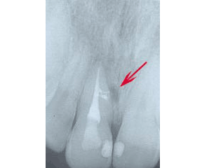
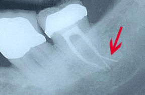
This is a different tooth that also had an accessory canal, in the area indicated by the red arrow. The prognosis for this tooth is also good, but as with most root canals, a periodic exam and x-ray are needed to monitor for any issues.

Root Canal Before

This is an example of a back tooth that developed a cyst (the dark area around the tip of the root). A root canal was performed and you can see some of the root canal filling material in the dark area, which represents an accessory canal. The prognosis for this tooth is good because the accessory canal was found and filled.

Root Canal After

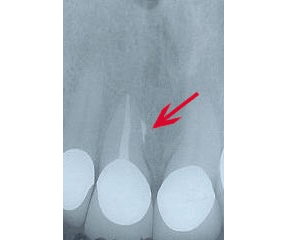
This is another example of an accessory canal in a back tooth. This canal,by the red arrow, was rather large. The prognosis for this tooth is also good because the accessory canal was found and filled.

Root Canal Before

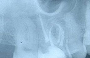
This is an x-ray of an upper molar with four canals, which is not very common. What makes the treatment of this tooth more difficult is the curvy nature of its canals.

Root Canal After

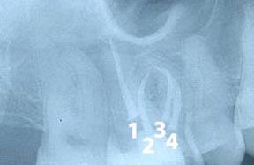
The four canals have been labeled on this x-ray. Canal #1 is very straight but the other three canals, especially #3, are curved. Each canal must be free of any nerve or blood tissue before it is filled to ensure a good prognosis.

Root Canal Before

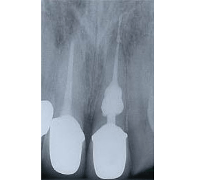
Most of the canals, where the nerve and blood vessels of the tooth reside, are narrow like the wick of a candle. Some canals are very wide, similar to this x-ray of a front tooth. This means there is less tooth structure surrounding the canal, which can indicate that the tooth is weaker.

Root Canal After

The tooth on the right is what we typically expect to see in regard to a normal root canal anatomy. This tooth has three separate canals. The tooth on the left has only a single canal, which is why the filling material appears so wide. The internal anatomy of each tooth is different, which makes each root canal procedure unique.

Root Canal Before

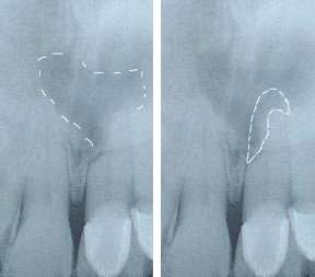
This is an x-ray of an upper front tooth. The root on this tooth is shorter than normal. The cells of the root of the tooth are being eaten away in a process that is called external resorption. This process usually occurs very slowly over a several months or years and is usually associated with some sort of trauma to the tooth. The dotted line on the left image above indicates an infection from the left tooth. The dotted line on the right image above represents that portion of the root of the tooth that has been lost due to the resorption.

Root Canal After

The treatment is a conventional root canal procedure, which may take one or two visits. This procedure is a last attempt at saving the tooth. The success of this procedure depends upon when the resorption is diagnosed and treated, the length of the root of the tooth, and the health of the surrounding bone. An annual exam with x-rays is the only way to monitor the prognosis of the tooth. In the case above, both teeth showed signs of resorption, which is why they were each treated with a root canal.

Root Canal Before

This is an x-ray of a back tooth, known as a molar, which was taken immediately after root canal treatment. The arrow is pointing to a dark area, where there is an infection.

Root Canal After

This x-ray was taken approximately one year after the root canal treatment. Notice that the dark area has filled in with bone, indicating that the root canal was successful. The large white area on the top portion of the tooth is a crown, which is required treatment for almost all back teeth that have a root canal.

Root Canal Before

The yellow arrow above indicates internal resorption. This occurs when the cells inside the tooth destroy the root from the inside out. This process usually happens very slowly over a matter of months or years and is usually associated with some sort of trauma to the tooth.

Root Canal After

The treatment for internal resorption usually takes a two or more appointments. First, a medicated filling is placed and then, after a few weeks, the medicated filling is removed and the procedure is completed in the conventional way. This type of condition requires annual examination by x-ray to check on the tooth. The prognosis is fair to poor, but the only other option is to extract the tooth.

Root Canal Before

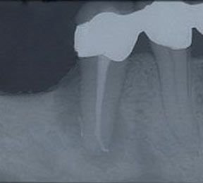
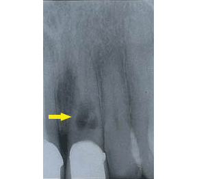
The dark area around the entire root of the tooth above indicates the presence of a large infection.

Root Canal After

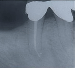
Conventional root canal treatment was performed and this is an x-ray of the tooth about six months later. You can see the dark infected area has disappeared and has been replaced by healthy bone structure. The prognosis for this tooth is excellent.


Learn More

  • Smile Gallery
  • Office Tour
  • Patient Education
  • Blog

Patient Education

Patient Education
  • Dental Services
    • Cleanings and Prevention
      • Bruxism and Mouthguards
      • Fluoride Treatments
      • Sealants
    • Cosmetic Dentistry
      • Bonding
      • Bridges
      • Crowns
      • Dentures
      • Fillings
      • Implants
        • Single Tooth
        • Multiple Teeth
        • All Teeth
      • Inlays and Onlays
      • Lumineers
        • Lumineers FAQ
        • Before and After
        • Perfect Smile
        • Lumismile
      • Teeth Whitening
      • Veneers
    • Gum and Bone
      • Bone Grafting
      • Cleanings and Prevention
      • Crown Lengthening
      • Frenectomy
      • Gum Tissue Graft
      • Perio Plastic Surgery
      • Ridge Augmentation
      • Scaling and Root Planing
      • Sinus Lift
    • Oral Surgery
      • Fractured Teeth
      • MDI Mini Dental Implants
      • Oral Cancer Exams and Biopsies
      • Pericoronitis
      • Wisdom Teeth
    • Orthodontics
      • ClearCorrect
      • Common Orthodontic Treatment
      • Craniofacial Orthodontics
      • Invisalign
      • Living with Braces
      • Myths and Fact About Orthodontics
      • Orthodontic FAQ
      • Orthodontic Glossary
      • Orthodontic Treatment for Children
      • Post Op Care of Extractions
      • Retainers
      • Six Month Smiles
        • Six Month Smiles FAQ
      • Space Maintainers
      • Surgical Orthodontics
      • Treatment for Adults
      • Treatment for Children
      • Two-Phase Orthodontic Treatment
      • Types of Appliances
      • Types of Braces
    • Pedodontics
      • Pulpotomy
      • Sealants
    • Root Canal
      • Knocked Out Tooth
      • Root Canal FAQ
      • Root Canal Surgery
      • Traumatic Injuries
    • Sedation Dentistry
    • TMJ
  • Patient Information
    • Dental Glossary
    • Development of the Teeth
    • FAQ
    • Fun Links for Kids
    • Patient Education
      • Acid Experiment
      • ANUG Trench Mouth
      • Bad Breath
      • Bruxism Tooth Grinding
      • Chemo and Your Mouth
      • Conditions of the Tongue
      • Dental X Rays
      • Denture Care
      • Dry Mouth
      • Fluoride Experiment
      • How to Brush
      • How to Floss
      • How to Read X Rays
      • Oncology Guide
      • Smokeless Tobacco
      • Tooth Anatomy
      • Tooth Sensitivity
      • Toothpaste Ingredients
    • Post Op Instructions
      • Crowns and Bridges Post Op
      • Dentures Post Op
      • Extractions Post Op
      • Fillings Post Op
      • Gum Surgery Post Op
      • Porcelain Veneers Post Op
      • Root Canal Post Op
      • Scaling and Root Planing Post Op
    • Teeth Trivia
    • Tips for Parents
    • Useful Dental Links
  • Smile Gallery
    • Bonding Gallery
    • Crowns Gallery
    • Dentures Gallery
    • Fillings Gallery
    • Implants Gallery
    • Orthodontic Gallery
    • Periodontal Gallery
    • Root Canal Gallery
    • Six Month Smiles Gallery
    • Veneers Gallery
    • Whitening Gallery
  • Technology
    • 3i Dental Implants
    • 3M Mini Dental Implants
    • AcquaBrite
    • Ankylos C/X Implants
    • BioHorizons
    • Brux-eze
    • CEREC One Appointment Crowns
    • ClearCorrect
    • Cone Beam Computed Tomography (CBCT)
    • Dash In-Office Whitening System by Discus Dental
    • DenOptix Digital X Rays
    • Dental Microscope
    • Dentrix Intra Oral Camera
    • DEXcam Intra-oral Camera
    • DEXIS Digital X Rays
    • DIAGNOdent
    • Discus Dental NiteWhite ACP Take-Home Whitening Sy
    • Discus NiteWhite ACP
    • E4D Same-Day Crowns
    • Florida Probe
    • Gendex Digital X-Ray
    • Gendex Intraoral Camera
    • Instrumentarium Panoramic X-ray
    • Invisalign
      • Invisalign Teen
    • Intraoral camera
    • Isolite Dryfield Illuminator
    • iTero Digital Impressions
    • Kodak Digital X Rays
    • Kor Whitening
    • LaserSmile
    • Lightyear Digital X Ray System
    • Lumineers
    • MagicFil
    • MDI Implants
    • Nitrous Oxide
    • Nobel Biocare
    • NTI System
    • Odyssey Diode Laser
    • Opalescense Boost
    • Opalescense Take Home
    • Opalescense Treswhite
    • OraVerse
    • Perio Protect
    • Pola Whitening for Life
    • Rembrandt Whitening
    • ScanX Digital X Ray
    • Schick Digital X Ray
    • 6 Month Smiles
    • Snap On Smile
    • SOPRO Intraoral Camera
    • Soredex Cranex D Panoramic X-ray
    • Straumann-Dental-Implants
    • Toothprints
    • Under Armour Mouthguards
      • Under Armour FAQ
    • VELscope
    • Vizilite Plus
    • The Wand
    • Waterlase
    • XDR Digital X Ray
    • Zoom Chairside
    • Zoom Take Home

Our Location

2607 Davie Boulevard
Ft. Lauderdale, FL 33312
(954) 587-7111

Map & directions

Dentist - Ft. Lauderdale, Llera Dental, 2607 Davie Boulevard, Ft. Lauderdale, FL 33312 (954) 587-7111

  • Home
  • Staff
  • Our Locations
  • Services
  • New Patients
  • Patient Education
  • Contact Us
  • Appointment Request
  • Website for Dentist Copyright © MH Sub I, LLC dba Officite
  • Disclaimer
  • Patient Privacy
  • Site Map